Hydrocephalus, etiology, types, & management.
Definition: – Spina bifida, which means “cleft spine,” is characterized by the incomplete development of the brain, spinal cord, and/or meninges. Etiology: – The exact cause of spina bifida remains a …
Definition: –
Spina bifida, which means “cleft spine,” is characterized by the incomplete development of the brain, spinal cord, and/or meninges.
Etiology: –
The exact cause of spina bifida remains a mystery. No one knows what disrupts complete closure of the neural tube, causing this malformation to develop. Suspect factors that cause spina bifida are multiple:-
• Genetic
• Nutritional – insufficient intake of folic acid—a common B vitamin—in the mother’s diet is a key factor in causing spina bifida and other neural tube defects. Prenatal vitamins typically contain folic acid as well as other vitamins.
• Environmental factors all play a role.
Types: –
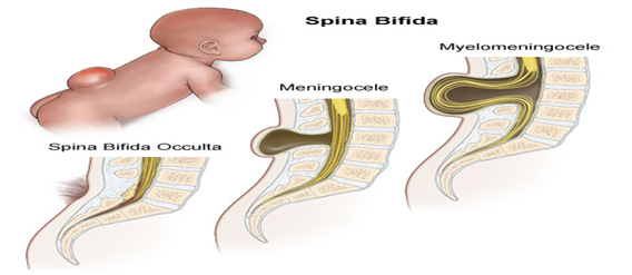
Spina Bifida Occulta: –
Occulta is the mildest and most common form in which one or more vertebrae are malformed. It indicates that a layer of skin covers the malformation, or opening in the vertebrae. This form of spina bifida, present in 10-20 percent of the general population, rarely causes disability or symptoms.
Spina Bifida Cystica: –
Closed neural tube defects make up the second type of spina bifida.It also called as spina bifida cystic. This form consists of a diverse group of defects in which the spinal cord is marked by malformations of fat, bone, or meninges.
There are mainly two types: –
Meningocele: –
spinal fluid and meninges protrude through an abnormal vertebral opening; the malformation contains no neural elements and may or may not be covered by a layer of skin. Some individuals with meningocele may have few or no symptoms while others may experience such symptoms as complete paralysis with bladder and bowel dysfunction.
Myelomeningocele: – the fourth form, is the most severe and occurs when the spinal cord/neural elements are exposed through the opening in the spine. The impairment may be so severe that the affected individual is unable to walk and may have bladder and bowel dysfunction.
Management: –
Surgical management- Meningocele involves surgery to put the meninges back in place and close the opening in the vertebrae. Myelomeningocele also requires surgery, usually within 24 to 48 hours after birth.
Performing the surgery early can help minimize risk of infection that’s associated with the exposed nerves and may also help protect the spinal cord from additional trauma.
During the procedure, a neurosurgeon places the spinal cord and exposed tissue inside the baby’s body and covers them with muscle and skin. Sometimes a shunt to control hydrocephalus in the baby’s brain is placed during the operation on the spinal cord.
Treatment doesn’t end with the initial surgery, though. In babies with myelomeningocele, irreparable nerve damage has already occurred and ongoing care from a multidisciplinary team of surgeons, physicians and therapists is usually needed. Babies with myelomeningocele may need further care for a variety of complications.


